FIGURE 2.

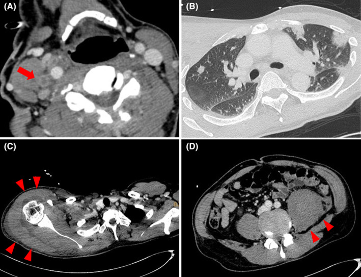
Computed tomography reveals (A) a thrombus of the right internal jugular vein (red arrow), (B) septic pulmonary embolisms, and (C, D) abscesses around the right shoulder and left psoas major muscle (red arrowhead).

Computed tomography reveals (A) a thrombus of the right internal jugular vein (red arrow), (B) septic pulmonary embolisms, and (C, D) abscesses around the right shoulder and left psoas major muscle (red arrowhead).