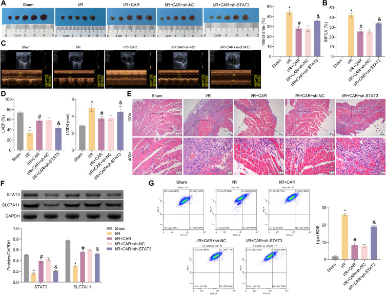
Figure 8.
CAR targeting STAT3 signaling inhibits MIRI-induced ferroptosis in mice. (A) The left panel displays illustrative images of myocardial tissues, while the right panel denotes the percentage of infarction size. (B) Measurement of the injury severity assessed through INF/LV quantification. (C) Illustrative echocardiograms captured at 4 h following I/R injury. (D) The ejection fraction of the left ventricle (LVEF) and diastolic diameter of the left ventricle (LVIDd) were shown. (E) Representative images of HE staining of myocardial tissues. (F) The expression of STAT3 and SLC7A11 was evaluated using Western blotting. (G) The lipid ROS level was assessed through flow cytometry. n = 3 per group. *P < 0.05 vs Sham group, #P < 0.05 vs I/R group, &P < 0.05 vs I/R+CAR+sh-NC group.

