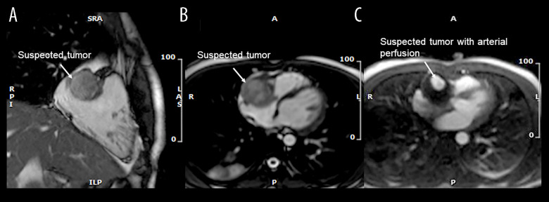
Figure 2.
Cardiac magnetic resonance. (A) 2-chamber view. A localized non-invasive suspected tumor (white arrow). (B) Transverse view. Suspected tumor (white arrow) compressing the tricuspid annulus. (C) Transverse view with dynamic perfusion. The suspected tumor (white arrow) with arterial perfusion.

