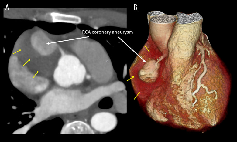
Figure 3.
Cardiac computed tomography. (A) Cross-sectional view illustrating the giant coronary artery aneurysm (CAA) on the right coronary artery (RCA) (white arrow) surrounded by the thrombosed part of the giant CAA yellow arrows). (B) Volume-rendered image illustrating the giant CAA on the RCA (white arrow) surrounded by the thrombosed part (yellow arrows).

