Fig. 1.
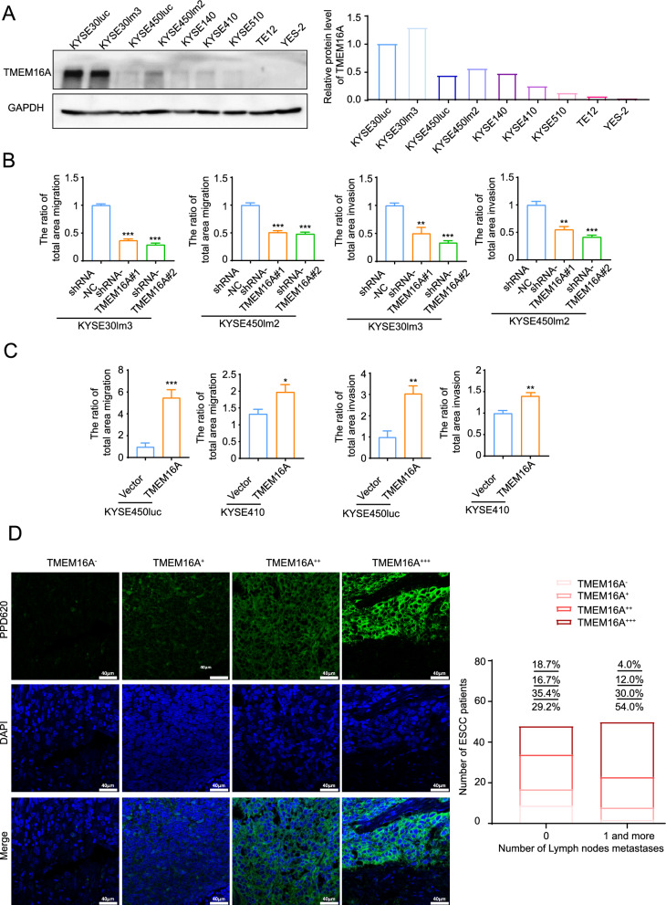
TMEM16A promotes ESCC cell motility and lymph node metastasis. A TMEM16A was more highly expressed in ESCC cells with high metastatic ability. Western blot (left) and grayscale scanning analysis (right) of TMEM16A level in ESCC cell lines. GAPDH was used as a loading control. B, C TMEM16A promotes ESCC cell migration and invasion. B Transwell chamber assays and Matrigel invasion assays for KYSE30lm3 and KYSE450lm2 cells after TMEM16A knockdown. C Transwell chamber assays and Matrigel invasion assays for KYSE450luc and KYSE410 cells overexpressing TMEM16A. D Fluorescence immunohistochemistry staining showed that the level of TMEM16A in ESCC tissues was significantly correlated with the degree of lymph node metastasis. The right figure shows the statistical graph of TMEM16A level and the number of lymph node metastasis in patients. The fluorescence values (MFI) of all samples were sorted numerically from smallest to largest based on the relative values of fluorescence intensity. High/low TMEM16A expression was distinguished according to MFI quartiles. Lower quartile Q1 was 31.145, denoted as " + ". Median Q2 was 38.506, and upper quartile Q3 was 43.821, denoted as " + + ". Samples with MFI greater than 43.821 were labeled as " + + + ". No detectable fluorescence intensity was denoted as “-”

