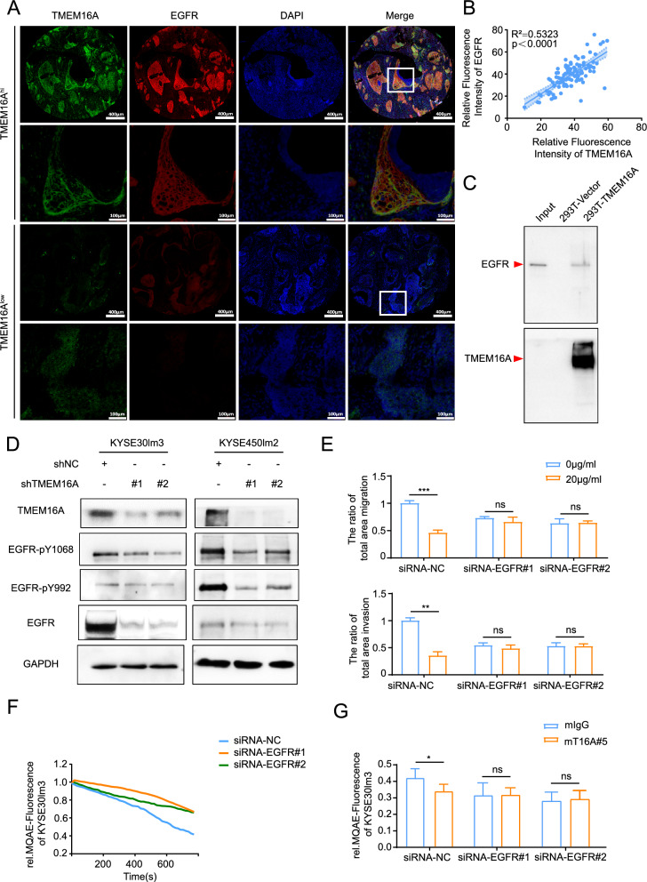
Fig. 3.
Mutual regulation between TMEM16A and EGFR. A, B Detection of TMEM16A and EGFR levels in tumors and adjacent normal tissues of 98 ESCC patients. A mIHC staining of tissues from patients with ESCC patients with fluorescent dye coupled to excitation light of 520 nm for the EGFR antibody, fluorescent dye coupled to excitation light of 650 nm for the TMEM16A antibody, and DAPI was used for staining nuclei. B The expression of TMEM16A and EGFR in ESCC tissues was positively correlated with each other. The staining results were statistically analyzed, and the spectral splitting and fluorescence semi-quantification were carried out after removing the background value of autofluorescence in the tissues. C Pull-down assay detected the interaction of TMEM16A with EGFR extracellular segment protein (aa 668–1210) in 293 T cells overexpressing TMEM16A. EGFR extracellular segment was used as Input. D Knockdown of TMEM16A down-regulated EGFR protein levels. E The inhibitory effect of mT16A#5 on migration and invasion of KYSE30lm3 cells was dependent on EGFR expression. F MQAE system assay showed that knockdown of EGFR was able to down-regulate the chloride channel activity of KYSE30lm3 cells. G. Effect of antibodies mIgG (20 μg/ml) and mT16A#5 (20 μg/ml) on the chloride channel activity in EGFR knockdown on KYSE30lm3 by the MQAE probe. **P < 0.01, ***P < 0.001, ns P > 0.05

