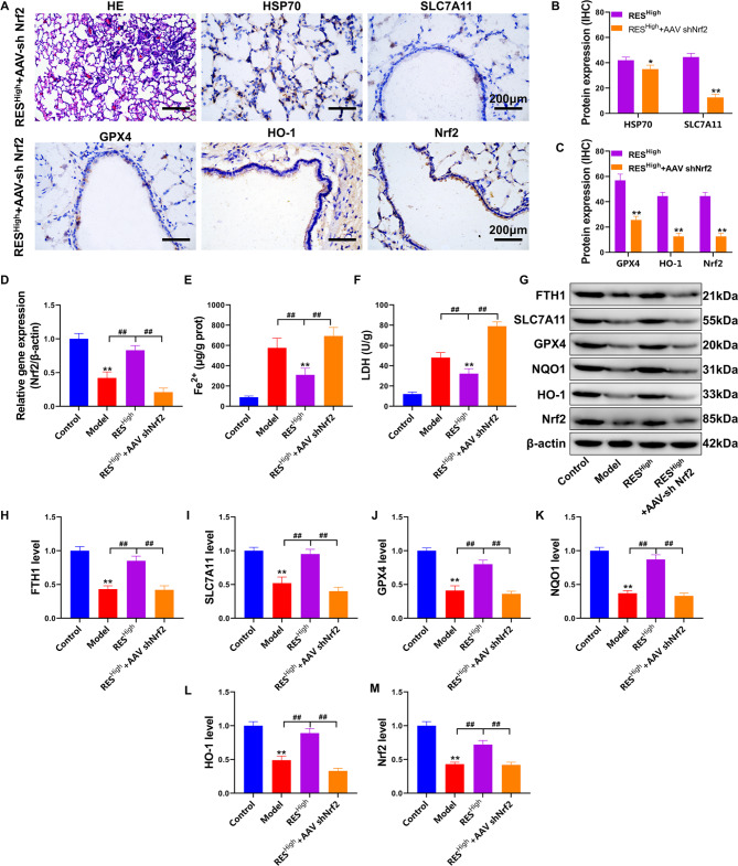
Fig. 6.
RES rescues pulmonary ferroptosis in HS rats via Nrf2. (A) Pathological examination of lung tissue via HE and IHC staining. (B-C) Quantitative statistics of IHC staining were analyzed and compared with those of the RESHigh group. (D) Detection of Nrf2 gene expression in lung tissue using RT-PCR. (E) Fe2+ levels in lung tissue. (F) LDH content detection. (G) FTH1, SLC7A11, GPX4, NQO1, HO-1, and Nrf2 protein expression levels, as determined using western blotting. (H–M) Quantification of the optical density values of protein bands determined using western blotting; **P < 0.01, *P < 0.05, compared with the control group; ##P < 0.01, #P < 0.05, the two groups connected were compared

