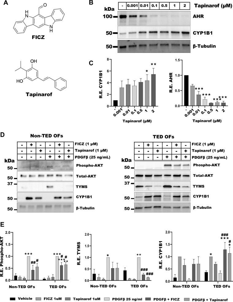
Figure 3.
AHR activation by FICZ or tapinarof attenuates PDGF signaling. (A) The 2D structures of FICZ and tapinarof. (B) TED OFs were treated with vehicle (DMSO) or tapinarof as indicated for 48 hours. Cells were analyzed by Western blot for AHR, CYP1B1, and β-tubulin (loading control). Tapinarof led to a dose dependent reduction in AHR levels and a dose dependent induction of CYP1B1 expression. (C) Quantification of Western blots for AHR and CYP1B1. (D) Non-TED and TED OFs were pretreated for 1 hour with FICZ (1 µM) or tapinarof (1 µM) followed by 48-hour stimulation with PDGFβ (25 ng/mL). Cells were harvested and analyzed by Western blot for phospho-AKT, total AKT, TYMS, CYP1B1, and β-tubulin (loading control). Phospho-AKT was normalized to total AKT, whereas other protein levels were normalized to β-tubulin. FICZ and tapinarof induced CYP1B1 levels. PDGFβ treatment induced phospho-AKT and TYMS, whereas FICZ and tapinarof mitigated these effects. Uncropped blots are presented in the Supplementary Data File. (E) Quantification of Western blots for phospho-AKT, TYMS, and CYP1B1. Vehicle (DMSO) treated cells served as control. The experiment was repeated in three non-TED and three TED OF strains with representative results shown. One-way ANOVA with Dunnett's multiple comparisons was used to analyze the data. * P ≤ 0.05, ** P = 0.005, *** P < 0.0008, and PDGF versus treatments: # P ≤ 0.05, ## P ≤ 0.005, ### P ≤ 0.0005.

