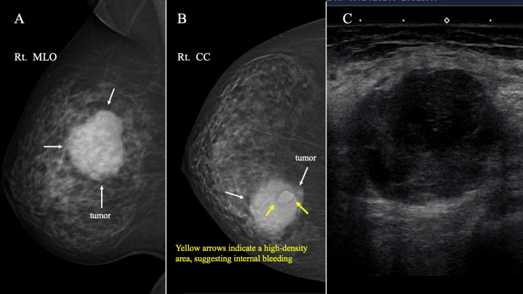
Figure 1. MG and US findings.
(A) MG shows a lobulated, high-density mass (white arrow) on medio-lateral oblique view. (B) A craniocaudal view of 3D-MG showed a lobulated mass (white arrow) with relatively high-density area (yellow arrow) on the internal chest wall side of the tumor. The yellow arrows indicate a high-density area, suggesting internal bleeding. (C) US findings. US revealed a hypoechoic, heterogeneous tumor with an unclear border.
MG: Mammography, US: Ultrasonography, 3D-MG: Tomosynthesis.

