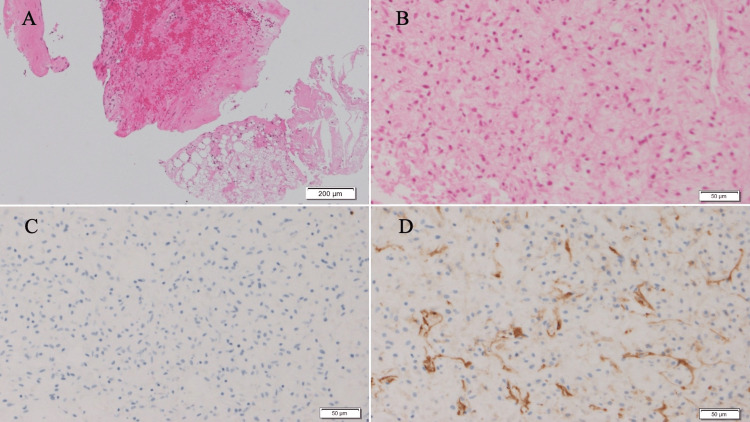
Figure 3. Pathological findings of CNB.
The fragmented breast tissue was stained with hematoxylin and eosin staining, which revealed (A) fibroedematous stroma with hemorrhage (scale bar: 200 μm) and (B) short spindle cells (scale bar: 50 μm). Immunohistological staining of the spindle cells showed (C) S-100 (-), (D) CD34 (+, focal) (scale bar: 50 μm).
CNB: Core needle biopsy, CD: Cluster of differentiation.

