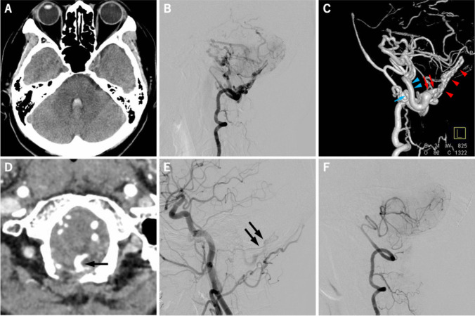
Fig. 2.
A 60-year-old female presented sudden onset of severe headache. The computed tomography (CT) image on admission showed the small amount of subarachnoid hemorrhage (SAH) and intraventricular hemorrhage (IVH) in the fourth ventricle (a). The lateral view of right vertebral angiogram (VAG) revealed high-flow dural arteriovenous fistula (dAVF) at foramen magnum region (FMR) with marked dilatation of posterior fossa veins (b). Left lateral view of 3-dementional rotational right VAG demonstrated shunted pouch on the surface of dura matter (red arrowhead) and the bridging vein (BV: red arrow) that works as a single drainer of dAVF and connected to vein of inferior cerebellar peduncle (blue arrowhead) and to posterior medial medullary vein (blue arrow) (c). The BV (arrow) that connecting FMR and posterior fossa veins was demonstrated by the source image of CT angiography (d). The lateral view of left carotid angiogram after endovascular transarterial embolization (TAE) of main feeders showed a small amount of residual shunt flow from the dural branches of ascending pharyngeal artery (e). The lateral view of right VAG obtained two weeks after surgical interruption showed the complete disappearance of high-flow FMR-dAVF (f)

