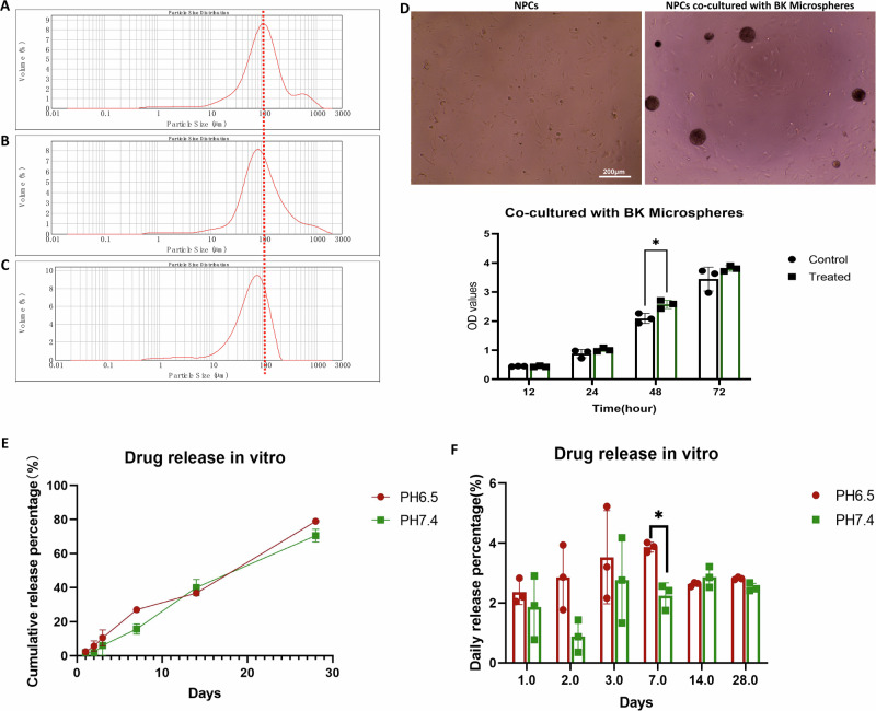
Fig. 6. Characterization, cell co-culture, and drug release of PLGA/BK microspheres.
A Particle size distribution of microspheres with an input of 200 mg of PLGA, with a median particle size of 95 μm. B Particle size distribution of microspheres with an input of 100 mg of PLGA, with a median particle size of 87 μm. C Particle size distribution of microspheres with an input of 50 mg of PLGA, with a median particle size of 60 μm. D Images of isolated cell groups, co-culture of microspheres and cells in a 96-well plate, and CCK-8 results for PLGA/BK microspheres co-cultured with primary rat NPCs for 3 days in vitro. E, F In vitro drug release of PLGA/BK microspheres in PBS with different pH. E Cumulative drug release from the microspheres. F Average daily drug release from the microspheres.

