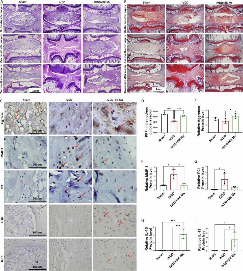
Fig. 8. Histological and immunohistochemical analysis of intervertebral discs post-surgery.
A HE staining of intervertebral disks 1 day, 1 month, and 2 months after surgery. B Safranin O-fast green staining of the intervertebral disc 1 day, 1 month, and 2 months after surgery. C IHC: Aggrecan, MMP-3, P21, IL-1β and IL-18 staining in intervertebral disks two months after surgery. Red arrows indicate positive cells. D PPP(Percentage of Positive Pixel) in the nucleus pulposus region. E Relative Aggrecan protein levels. F Relative MMP-3 protein levels. G Relative P21 protein levels. H Relative IL-1β protein levels. I Relative IL-18 protein levels. To assess the statistical differences between various groups, we employed one-way analysis of variance (ANOVA) followed by Tukey’s post hoc test for multiple comparisons (*: p < 0.05, **: p < 0.01, ***: p < 0.001, ****: p < 0.0001). At each time point, each group consisted of three rats (n = 3).

