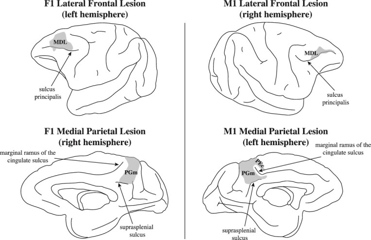
Fig. 4.

Location of crossed unilateral lesions in macaque monkeys F1 and M1. Note that the frontal lesion in monkey F1 involves the entire mid-dorsolateral prefrontal region (areas 46 and 9/46). The frontal lesion in monkey M1 is restricted to the anterior part of the mid-dorsolateral prefrontal region, sparing its posterior part. Note that monkey F1 demonstrated the more severe impairment (see Fig. 5). On the medial hemisphere of monkey F1, the lesion was restricted to the precuneal medial parietal area PGm. On the medial hemisphere of monkey M1, the lesion included area PGm, but extended above the marginal ramus of the cingulate sulcus to include a small portion of area PEc. Abbreviations: MDL, mid-dorsolateral prefrontal region; area PGm on the medial parietal cortical region, i.e. the precuneus.
