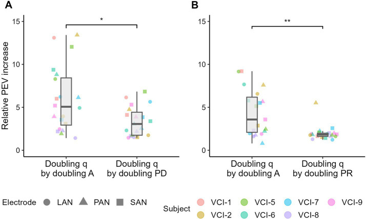
Figure 6.
PEV, charge (q) based, comparison between (A) the stimulation amplitude (A) and phase duration (PD) and (B) between the stimulation amplitude and pulse rate (PR). Comparison is made by doubling the delivered charge by doubling the amplitude, phase duration and stimulation rate. In each figure, the paired samples have the same delivered charge. Relative PEV increase was calculated by dividing the doubled charge PEV by the initial PEV. Boxplots indicate the median and interquartile ranges of the relative PEV increase. Paired two-sided Wilcoxon signed-rank tests were performed, with * indicating p < 0.001. Note that the data was obtained in two experiments (amplitude versus phase duration, and amplitude versus pulse rate), which explains the difference between the data in the two “doubling q by doubling A” boxplots.

