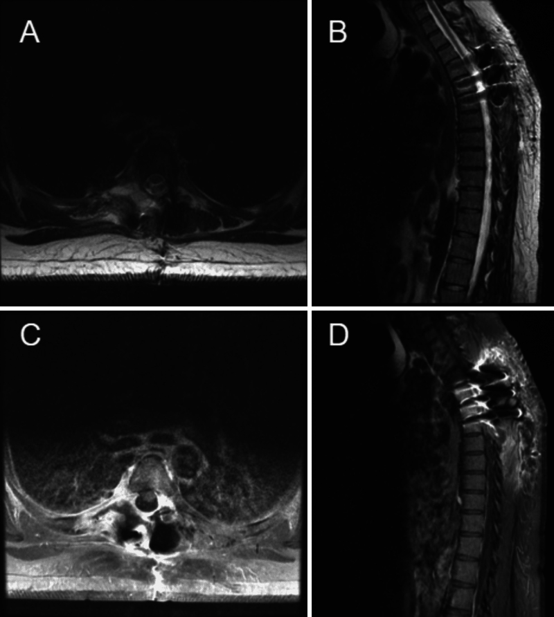
FIG. 4.
One-month postoperative MRI. A: Axial T2-weighted image at the level of T5, without obvious residual of the previous fluid-filled cystic lesion. B: Sagittal T2-weighted image showing postsurgical changes and resolution of the previous fluid-filled cystic lesion abutting the pleura. C: Axial postcontrast T1-weighted image at the level of T5 showing no enhancing remnant of the previous lesion near the vertebra. D: Sagittal postcontrast T1-weighted image without enhancing remnant of the previous lesion on the pleura.

