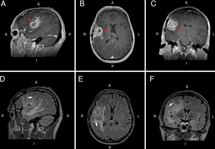
FIG. 1.
Preoperative sagittal (A), axial (B), and coronal (C) contrast T1-weighted MRI slices indicate a heterogeneously enhancing mass (red arrows) in the right frontoparietal operculum. Corresponding preoperative FLAIR MRI slices (D−F) indicate peritumoral edema extending toward the cortical surface (white arrows).

