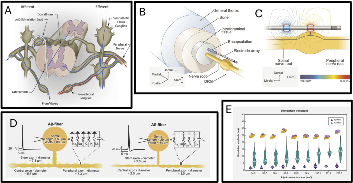
FIGURE 8.
(A) Diagram of dorsal root ganglion stimulation electrode implanted in foramen interbod (Sverrisdottir et al., 2020). (B) Schematic diagram of three-dimensional finite element model of dorsal root nerve stimulation (Graham et al., 2022a). (C) Isopotential lines of the extracellular voltages generated by bipolar DRGS calculated from the FEM (Graham et al., 2022a). (D) Multi-compartment models of two types primary sensory neurons (Bhowmick et al., 2024). (E) The distribution of activation thresholds of Aβ- and Aδ-fibers generated by the various Injectrode geometries with the mean values inset and the corresponding Injectrode geometry at the top of each violin plot (Bhowmick et al., 2024).

