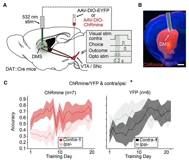
Figure 4. Stimulating DMS DA terminals at the onset of contralateral stimulus presentation improves side-specific performance.
(A) Schematic of the optogenetic stimulation of DMS DA terminals. Mice either expressed ChRmine or a control construct in DA neurons. DA terminals in the DMS were optogenetically stimulated unilaterally (532 nm, 0.2 s burst duration, 5 ms pulse width, 20 Hz pulses, ~0.25 mW) at the onset of the contralateral stimulus presentation throughout training.
(B) Example histology image of optical fiber location and terminal expression of ChRmine-mScarlet. Scale bar, 900 μm.
(C) Comparison of performance for contralateral and ipsilateral stimulus trials in control (n = 7, left) and ChRmine (n = 6, right) mice. Lines and shading represent mean ± SEM. *p < 0.05 for cohort (ChRmine/YFP) and side (contra/ipsi) interaction in three-way ANOVA with cohort (ChRmine/YFP), day, and side (contralateral/ipsilateral) as factors (see Table S2 for model details and full results).
See also Figures S1 and S2 and Table S2.

