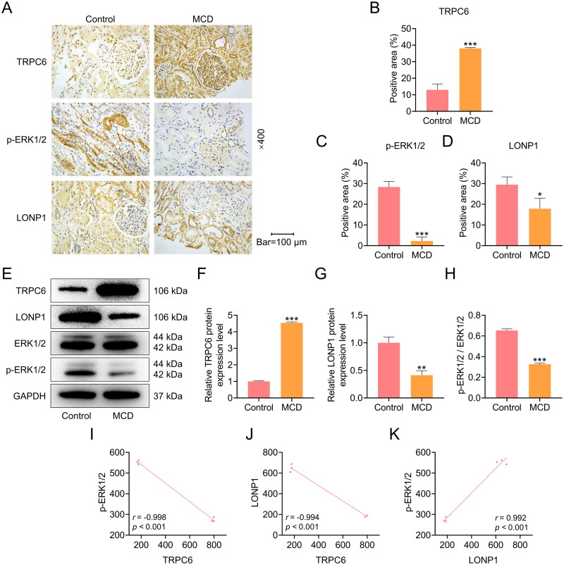
Figure 1.
The protein expressions of TRPC6, LONP1 and p-ERK1/2 in the kidney tissues of MCD patients.
(A-D) The expressions of TRPC6, p-ERK1/2, and LONP1 in the kidney tissues of MCD patients were detected by immunohistochemistry (magnification: ×400, scale bar = 100 µm). (E-H) The protein expressions of TRPC6, p-ERK1/2, ERK1/2 and LONP1 in the kidney tissues were measured by Western blot. GAPDH was used as a loading control. (I) The correlation between TRPC6 and p-ERK1/2 (r = -0.998, p < 0.001). (J) The correlation between TRPC6 and LONP1 (r = -0.994, p < 0.001). (K) The correlation between LONP1 and p-ERK1/2 (r = 0.992, p < 0.001). The data are presented as the mean ± standard deviation of three independent experiments; **p < 0.01, ***p < 0.001 vs. Control. Abbreviation: TRPC6, transient receptor potential cation channel subfamily C member 6; GAPDH, glyceraldehyde-3-phosphate dehydrogenase; LONP1, Lon peptidase 1; ERK, extracellular signal-regulated kinase; p-ERK, phosphorylated ERK.

