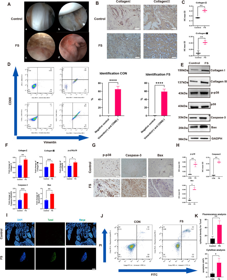
Fig. 3.
The FS tissue was successfully collected and digested into FS synovium fibroblasts. Besides phosphorylation level of p38 was higher and cell apoptosis was more active in the FS group. A The arthroscope view of Control (a,b) and FS(c,d) patient: the capsular was more thicken and the synovium was hyperemia and inflammation infiltrated. B Representative Collagen I and Collagen III immuno-stained tissue sections in control and FS tissue. Scale Bar: 50 μM. C The statistics analysis of (B): the mean optical density (mean OD) of Collagen I and Collagen III in the control and FS tissues (n = 3). D The representative flow cytometry results of synovial fibroblasts and the statistic results (n = 3). E, F The western blot analysis of Collagen I, Collagen III, p-p38, Caspase-3 and Bax. The statistic results (n = 3) confirmed that the above proteins were higher expressed in the FS group, which were corresponding with the IHC results. *P < 0.05; **P < 0.01; *** P < 0.001; **** P < 0.0001. G Representative p-p38, Caspase-3 and Bax immuno-stained tissue sections in control and FS tissue. Scale Bar: 50 μM. H The statistics analysis of (A): the mean optical density (mean OD) of p-p38, Caspase-3 and Bax in the control and FS tissues (n = 3). *P < 0.05; **P < 0.01; *** P < 0.001; **** P < 0.0001. I, J, K The cell apoptosis was more active in FS group based on the Tunel staining (I) and flow cytometry analysis (G). Scale Bar: 200 μM. *P < 0.05; **P < 0.01; *** P < 0.001; **** P < 0.0001

