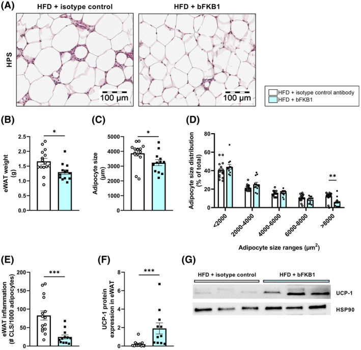
FIGURE 2.

Adipocyte size and adipose tissue inflammation are reduced by bFKB1. Representative histological photomicrographs of HPS‐stained epididymal white adipose tissue (eWAT) (A), epididymal WAT weight (B), average adipocyte size (C), adipocyte size distribution as percentage of total adipocytes (D), number of crown‐like structures (CLS) per 1000 adipocytes (E), uncoupling protein 1 (UCP1) protein expression in epididymal WAT normalized to HSP90 expression (F), and representative images of the Western blot bands (G). Values are presented as mean ± SEM for n = 15 mice on HFD treated with isotype control antibody and n = 13 mice on HFD treated with bFKB1. *p < .05, **p < .01, ***p < .001 bFKB1 versus isotype control antibody.
