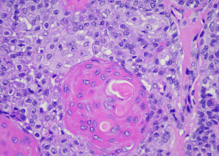
Figure 3. SEDC at high (400x) magnification showing enlarged pale squamoid cells, large cells with abundant eosinophilic cytoplasm, and few smaller basaloid cells surrounding well-formed small ductal lumen and larger keratinizing cystic spaces. The nuclei appear atypical and moderately pleomorphic with irregular nuclear contours and prominent nucleoli.
SEDC, squamoid eccrine ductal carcinoma

