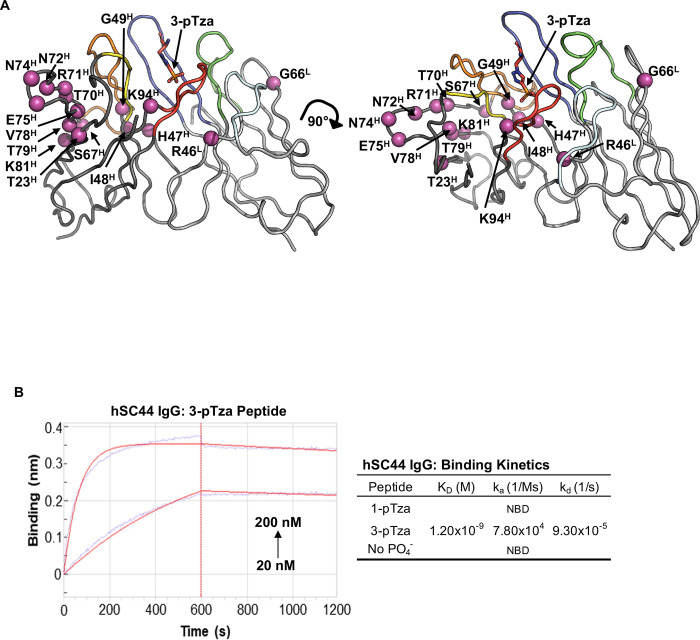
Fig. 1. Humanization of the rabbit (rSC44) antibody.
(A) Structure of rSC44 (PDB ID: 6X1V) in complex with a 3-pTza containing peptide (3-pTza shown as sticks colored in red) was used to guide humanization. The VH and VL domains are colored dark and light grey respectively. The CDRs are colored as follows: H3, red; H2, orange; H1, yellow; L3, blue; L2, cyan; L1, green. Residues in important framework positions, including known Vernier Zone residues are colored magenta and represented as spheres highlighting their Cα position. Residue numbering is in accordance with Kabat nomenclature. (B) Biolayer Interferometry (BLI) data for hSC44 IgG binding to 3-pTza, 1-pTza and a non-phosphorylated (no PO4−) peptide. The table shows the binding constants determined from BLI traces, and sample traces are shown for hSC44 binding to the immobilized 3-pTza peptide. NBD indicates no binding detected.

