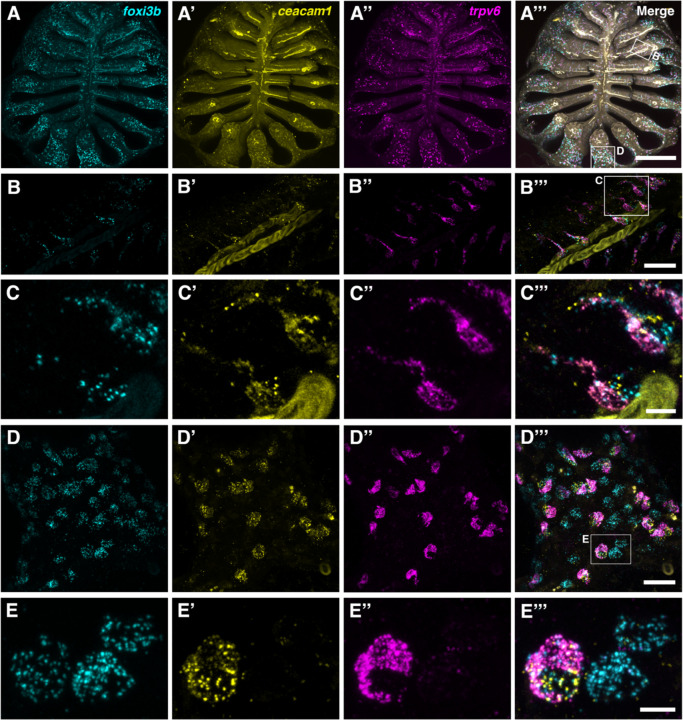
Figure 3. The adult zebrafish olfactory epithelium contains three distinct subtypes of ionocytes.
(A–A’’’) Overview of a dissected adult olfactory rosette. Maximum intensity projections of an Airyscan2 confocal image of HCR RNA-FISH for foxi3b (A), ceacam1 (A’), trpv6 (A’’), and merged signals (A’’’). Scale bar: 200 μm. (B–B’’’) Enlargement of the boxed region ‘B’ in A’’’, within the central (sensory) zone of the olfactory rosette. Maximum intensity projections of selected Airyscan2 confocal slices; HCR RNA-FISH for foxi3b (B), ceacam1 (B’), trpv6 (B’’), and merged signals (B’’’). Pairs of elongated ionocytes with cell bodies located deep in the epithelium are visible. (The yellow stripe running through the image is autofluorescence from a blood vessel.) Scale bar: 20 μm. (C–C’’’) Enlargement (maximum intensity projection of selected z-slices) of boxed region in B’’’, featuring two ionocyte pairs: HR-like ionocytes expressing ceacam1 (yellow) and foxi3b (cyan), adjacent to NaR-like ionocytes expressing trpv6 (magenta) and a low level of ceacam1. HCR RNA-FISH for foxi3b (C), ceacam1 (C’), trpv6 (C’’), and merged signals (C’’’). Scale bar: 5 μm. (D–D’’’) Enlargement of boxed region ‘D’ in A’’’, within the peripheral (non-sensory, multiciliated) zone of the olfactory rosette. Maximum intensity projections of selected Airyscan2 confocal z-slices; HCR RNA-FISH for foxi3b (D), ceacam1 (D’), trpv6 (D’’), and merged signals (D’’’). Both paired and solitary ionocytes are present. Scale bar: 20 μm. (E–E’’’). Enlargement (maximum intensity projection of selected z-slices) of the boxed region in D’’’, featuring one HR-like/NaR-like ionocyte pair, and two NCC-like ionocytes. An HR-like ionocyte, expressing ceacam1 (yellow) and foxi3b (cyan), sits adjacent to an NaR-like ionocyte expressing trpv6 (magenta) and a lower level of ceacam1. The NCC-like ionocytes express foxi3b (cyan) but not ceacam1 or trpv6. Ionocytes near the periphery of the rosette were rounded in shape. Scale bar: 5 μm.

