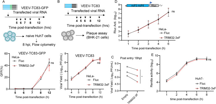
Fig 3. TRIM32 does not influence post-entry stages of the VEEV infection cycle.
A. Upper: schematic of post-entry assay by transfecting VEEV-TC83-GFP infectious RNA on HeLa-empty or HeLa-TRIM32 cells. Bottom: the supernatants were harvested at the indicated time points post-transfection and used to infect naïve HeLa cells. Infectivity of progeny virus was determined by flow cytometry. B. Upper schematic of post-entry assay by transfecting VEEV-TC83 infectious RNA on HeLa-Fluc or HeLa-TRIM32 cells. Bottom: the supernatants were collected at the indicated time points post-transfection. The progeny virus in supernatants were determined by plaque assay on BHK-21 cells. C. HeLa-Fluc or HeLa-TRIM32 cells were transfected with VEEV-TC83 infectious RNA for 18 hrs, the supernatants were collected and the progeny virus in supernatants were determined by plaque assay on BHK-21 cells. 18hpt, 18 hours post-transfection. Each point represents a biological replicate, and the paired biological replicate is connected by a straight line. D. HeLa-Empty or HeLa-TRIM32 cells were transfected with VEEV-TC83-Renilla luciferase (Rluc) replicon RNA. The cells were harvested at indicated time points, and Rluc activity was quantified. E. VEEV-TC83-Renilla luciferase (Rluc) replicon assay on Huh7-Empty or Huh7-TRIM32. Three independent biological replicates were carried out for each experiment. Statistical significance was determined by 2-way ANOVA with Sidak’s multiple comparisons test for A, B, D, E, ns, no significant. A paired t-test was used for C (*P<0.05).

