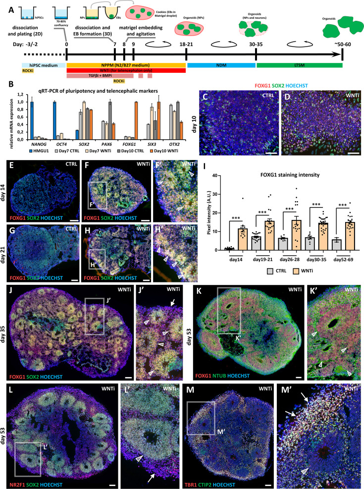
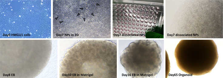
Figure 1. Hybrid 2D/3D protocol for fast and reproducible generation of human cortical organoids.
(A) Schematic of the hybrid 2D/3D method for generating telencephalic/cortical human organoids in vitro, using a triple inhibition of TGFβ, BMP, and WNT pathways (SB-431542, 5 µM; LDN-193189, 0.25 µM; XAV-939, 2 µM). On day 7, cells are dissociated and re-aggregated in 96-well plates. One day later, embryoid bodies (EBs) are embedded in Matrigel droplets (10 µL per droplet containing 1–4 EBs). These droplets, termed ‘cookies’, are then cultured in spinning bioreactors. (B) Real-time qRT-PCR analysis quantifying pluripotency markers (NANOG, OCT4) and telencephalic neural progenitor (NP) markers (SOX2, PAX6, FOXG1, SIX3, and OTX2) in undifferentiated HMGU1 hiPSCs and in day7 and day10 control (CTRL) and WNT-inhibited (WNTi) samples, as indicated. n=2 culture wells per condition, pooled prior to RNA extraction. (C,D) Immunostaining for FOXG1 (red) and SOX2 (green) in day10 2D neural cultures under control (CTRL) conditions (C) or following WNT inhibition (WNTi) (D). (E–I) Immunostaining for FOXG1 (red) and SOX2 (green) in day14 (E-F’) and day21 (G-H’) organoids under CTRL or WNTi conditions, as indicated. White arrowheads in high-magnification images point to neural progenitor (NP) rosettes. The graph (I) shows quantification of FOXG1 pixel intensity in CTRL and WNTi samples across time points. n≥7 sections from n≥4 organoids from n=2 independent batches (except day52-69 CTRL sample, n=2 sections from 1 batch). (J,J’) FOXG1 (red) and SOX2 (green) immunostaining in day35 WNTi organoids. White arrowheads in high-magnification images indicate NP neural rosettes, while arrows highlight differentiating neurons surrounding the rosettes. (K-M’) Immunostaining for FOXG1 (red) and NTUB (green) (K, K’), NR2F1 (red) and SOX2 (green) (L, L’), and TBR1 (red) and CTIP2 (green) (M, M’) in day53 WNTi organoids. High-magnification images highlight FOXG1+ SOX2+ NR2F1+ NP rosettes/neuroepithelia (K-L’; indicated by white arrowheads) surrounded by TBR1+ CTIP2+ NR2F1+ differentiating cortical neurons (L’-M’; indicated by white arrows). Scale bars: 100 µm.


