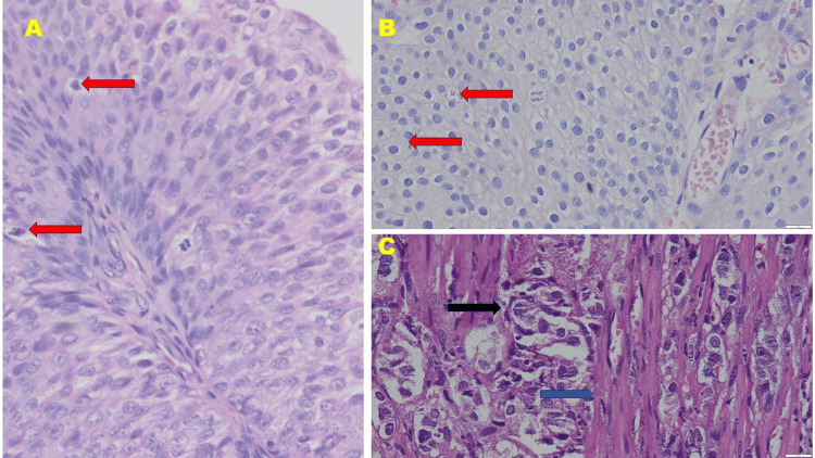
Figure 1. Histomorphology of (A) low-grade papillary urothelial carcinoma, (B) high-grade papillary urothelial carcinoma, and (C) invasive urothelial carcinoma (hematoxylin and eosin stain, 400× magnification) .
Note the atypical mitotic figures (shown by red arrow) in (A) low-grade papillary urothelial carcinoma and (B) high-grade papillary urothelial carcinoma. Also, note the invasion of malignant urothelial cells (black arrow) between the muscular layer (blue arrow) in (C) invasive urothelial carcinoma

