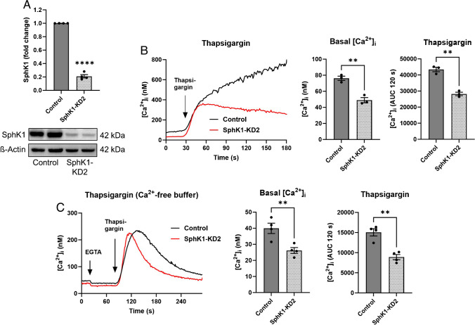
Fig. 2.
Ca2+ homeostasis in EA.hy926 cells with SphK1 knockdown, cell line 2 (SphK1-KD2). A Western blot analysis of SphK1 expression. Shown is a representative blot with duplicate samples, and the mean of four independent experiments performed in duplicates (means ± SEM; ****p < 0.0001 in one sample t-test). B, C Basal [Ca2+]i levels and Ca2+ increases induced by 1 µM thapsigargin were measured in fura-2-loaded cells in the presence (B) or absence (C) of extracellular Ca2+. Shown are representative traces of [Ca2+]i and means of 3 (B) or 4 (C) independent experiments. The response to thapsigargin was analyzed by measuring the [Ca2+]i increase above baseline (area under the curve, AUC) for 120 s after addition of thapsigargin (AUC 120 s). In C, 50 µM EGTA was added to the Ca2+-free buffer ~ 1 min before addition of thapsigargin (means ± SEM; **p < 0.01 Student’s t-test)

