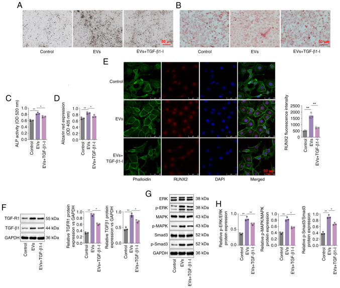
Figure 4.
DPSC-EVs may induce the osteogenic differentiation of HERS cells by activating TGF-β1/ERK signaling. (A) ALP staining was performed to evaluate the osteogenic ability of HERS cells. (B) Alizarin red staining was performed to evaluate the osteogenic ability of HERS cells. (C) ALP activity was detected in the HERS cells. (D) Alizarin red expression was detected in HERS cells. (E) Immunofluorescence staining was conducted on HERS cells to monitor the expression of RUNX2. (F) The expressions of TGF-β1 and TGFR1 in HERS cells were determined by Western blotting. (G) The activity of ERK/MAPK/Smad signaling in HERS cells were determined by Western blotting. (H) The expressions of MAPK, ERK, Smad3 and p-Smad3 were increased in the DPSC-EVs group compared with control group. The data from three independent experiments are presented as the mean ± SD; *P<0.05 and **P<0.01. DPSC, dental pulp stem cell; EV, extracellular vesicle; HERS, Hertwig's epithelial root sheath; p, phosphorylated; RUNX2, runt-related transcription factor 2; ALP, alkaline phosphatase.

