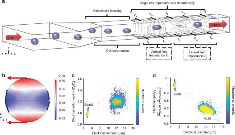
Fig. 1. Principle of the single-cell electro-mechanical shear flow deformability cytometer.
a Principle of the electro-mechanical cytometer. Cells suspended in a viscoelastic buffer flow along a microfluidic channel within which there are two sets of microelectrodes. One set measures the cell volume (Z1) whilst the second set measures cell deformation along the direction of flow (Z2). Cells are focused into the centre of the channel by the viscoelastic suspending fluid that is used to create the shear stress. b shows the shear stress on an undeformed sphere in a viscoelastic fluid (0.5% w/v methyl cellulose, 0.015 Pa·s, density 1005 kg/m3, flow rate 10 µl/min, particle radius 6 µm), see ESI for further details. Arrows indicate direction of the local force. Density plots of electrical deformability (c) and electrical opacity (d) as a function of electrical diameter for HL60 cells (n = 2000) at a flow rate of 10 μl/min. 5 µm rigid beads included as reference particles in both cases

