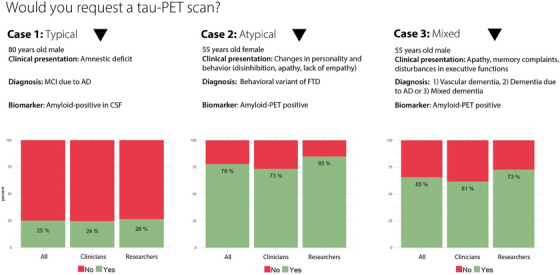
FIGURE 3.

Responses to three clinical case vignettes. Proportions of respondents who would request a tau‐PET scan in a typical AD patient (case 1), a patient with an atypical presentation (case 2), and a patient with suspected mixed pathology (case 3). AD, Alzheimer's disease; amyloid‐PET, amyloid positron emission tomography; CSF, cerebrospinal fluid; MCI, mild cognitive impairment; FTD, frontotemporal dementia; tau‐PET, tau positron emission tomography.
