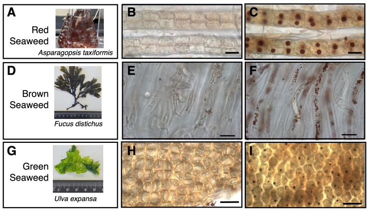
Figure 1. Detection of H 2 O 2 -containing organelles in red, brown, and green seaweeds.
A, D, and G, snapshots of seaweed organisms used in this study - red ( Asparagopsis taxiformis ), brown ( Fucus distichus ), and green ( Ulva expansa ), respectively. Fucus distichus and Ulva expansa are imaged with a ruler . B, E, H, as negative controls, 10 mM ascorbic acid was added to quench out H 2 O 2 before DAB staining in Asparagopsis taxiformis , Fucus distichus , and Ulva expansa , respectively. C, F, I, DAB staining to detect H 2 O 2 in Asparagopsis taxiformis , Fucus distichus , and Ulva expansa , respectively. Scale bar = 30 µm

