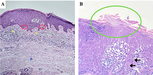
Figure 2.

(A) Histopathological examination (H&E, 100x) revealed vacuolar alteration (red circles), lymphohistiocytic infiltrates on dermoepidermal junction (yellow asterisks), and dermal solar elastosis (blue asterisk). (B) At higher magnification (H&E, 400x), a parakeratotic column with hypogranulosis (green circle) and few dyskeratotic cells (black arrows), resembling a cornoid lamella, was observed at the edge of the biopsy specimen.
