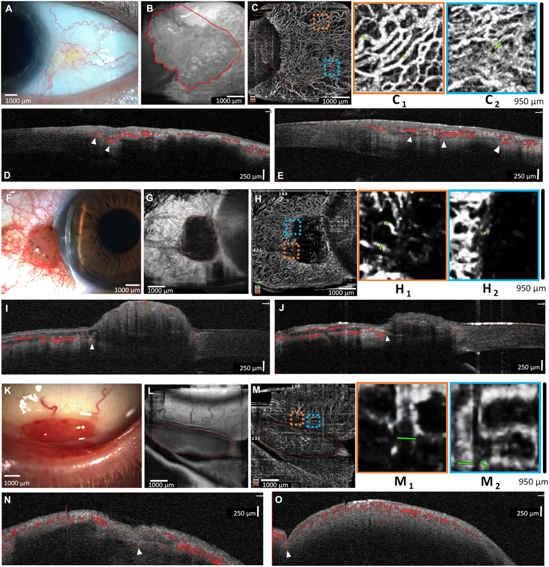
Fig. 2.

Representative images from malignant lesions. A) Slit-lamp photograph of a 58-year-old male with a conjunctival intra-epithelial neoplasia (CIN) of left eye. B) En face AS-OCT shows the lesion borders. C) AS-OCTA shows a highly vascularized conjunctiva, highlighting dilated peri-lesional vessels (C1 and C2). D and E) AS-OCT B-lines show the intra-lesional vessels are highly connected to the deep conjunctival and episcleral vessels at the lesion base (arrowheads). F) Slit-lamp photograph of a 59-year-old male with conjunctival melanoma of the right eye. G) En face AS-OCT shows lesion borders. H) AS-OCTA shows many peri-lesional vessels (highlighted in H1 and H2) with scant intra-lesional vessels. I and J) AS-OCT B-lines show superficial conjunctival vessels overlying lesion with deep episcleral branches entering lesion base (arrowheads). K) Slit-lamp photograph of a 64-year-old male with bilateral lymphoma in the inferior fornix. L) En face AS-OCT shows the lesion borders. M) AS-OCTA shows many peri-lesional vessels (highlighted in M1 and M2) and fine vessels overlying the lesion. N and O) AS-OCT B-lines highlight deep episcleral vessels entering the lesion base (arrowheads). All malignant lesions were biopsy proven.
