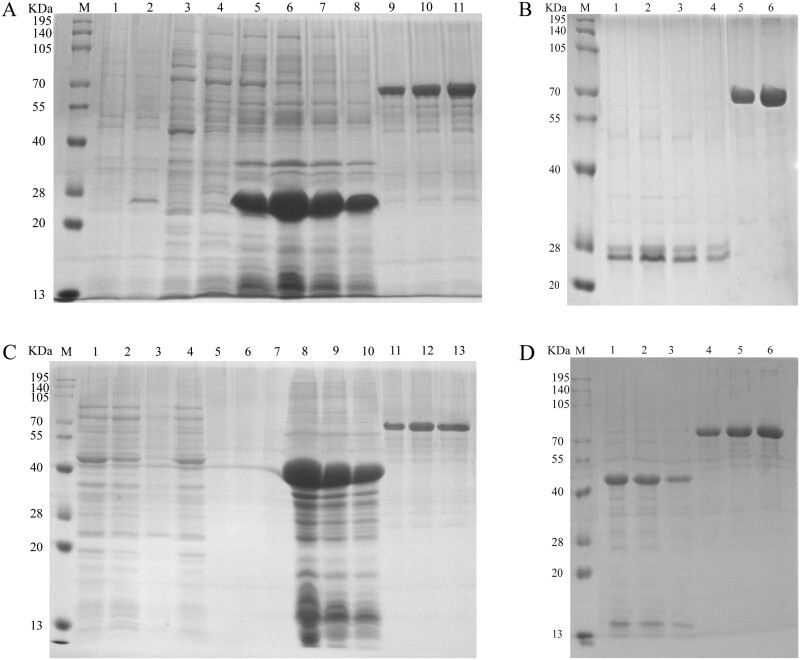
Fig. 7.
Purification of Cellulase-28a and GcEGaseZ7-28a. Note: M: Molecular weight of protein marker, size marked on the left; A) 1–2: Uninduced and induced expression bacteria of Cellulase-28a; 3–4: The fragmented supernatant of Cellulase-28a; 5–8: Undiluted, 5-fold dilution, 10-fold dilution, and 20-fold dilution of Cellulase-28a precipitates after fragmentation; 9–11: 0.2 mg/ml, 0.3 mg/ml, and 0.4 mg/ml of BSA. B) 1–4: Undiluted, 5-fold dilution, 10-fold dilution, and 20-fold dilution of Cellulase-28a dissolved in 8M urea and precipitated after fragmentation; 5–6: 0.2 mg/ml and 0.4 mg/ml of BSA. C) 1–3: Untreated, the supernatant of GcEGaseZ7-28a purified by His tag and the eluate of the supernatant of GcEGaseZ7-28a fragmented and purified by His tag; 4–7: Untreated, 5-fold dilution, 10-fold dilution, and 20-fold dilution after dialysis and concentration of the eluted protein obtained from the supernatant of GcEGaseZ7-28a purified by His tag; 8–10: 5-fold dilution, 10-fold dilution, and 20-fold dilution of GcEGaseZ7-28a precipitates after fragmentation; 11–13: 0.2 mg/ml, 0.3 mg/ml, and 0.4 mg/ml BSA. D) 1–3: 5-fold dilution, 10-fold dilution, and 20-fold dilution of GcEGaseZ7-28a dissolved in 8M urea and precipitated after fragmentation; 4–6: 0.2 mg/ml 0.3 mg/ml and 0.4 mg/ml BSA.

