Figure 2. a-d.

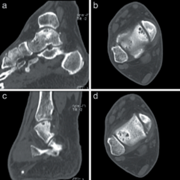
Preoperative computed tomography images. Preoperative (a) sagittal and (b) axial computed tomography images of OLT on medial talar dome. (c) Sagittal and (d) axial computed tomography images of OLT on lateral talar dome.

Preoperative computed tomography images. Preoperative (a) sagittal and (b) axial computed tomography images of OLT on medial talar dome. (c) Sagittal and (d) axial computed tomography images of OLT on lateral talar dome.