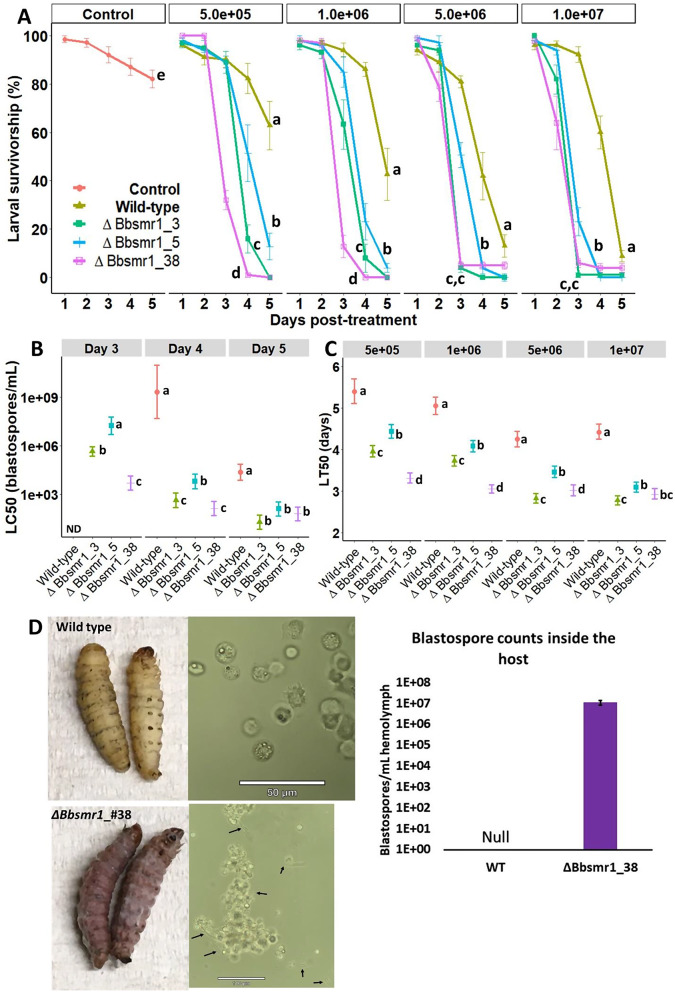
Fig. 4.
B. bassiana mutants display greater and faster killing activity against the greater wax moth compared with the wild-type strain. A) Survival probability of G. mellonella following exposure to crescent concentrations (5 × 10⁵ to 1 × 10⁷ blastospores/mL) of different knockout mutants (ΔBbsmr1) compared with the wild-type strain. Lettering indicates statistical differences based on the log-rank test (P < 0.05) for comparison between survival curves within each fungal concentration. Survival curves with means (± SE) represent data from five replicates (n = 100–140 insects/concentration/strain) derived from two independent bioassays. Lettering represents statistical differences (P < 0.05) based on a log-rank test comparing the Kaplan-Meier survival curves. Natural mortality in controls were attributed to unknown causes with dead larvae appearing all black and putrefied. B) LT50 (± 95% CI) values across different fungal concentrations. ND = not determined due to mortality level did not reach 50%. C) LC50 (± 95% CI) values across different time intervals following exposure to fungal treatments. The LC50 dose for untreated G. mellonella was fixed at zero and reported for all blastospore concentrations for comparison. Lettering of LT50s and LC50s indicates statistical differences based on the non-overlapping of their 95% CIs (P < 0.05). D) Microscopic observation of fungal development and insect immune responses to the WT and mutant strains at different times after cuticle-contact infection. No evidence of aggregation of hemocytes in the hemolymph from wild-type-infected larvae, whereas at within 3 days after exposure to fungal treatments of B. bassiana mutants, blastospores (indicated by arrows) appeared in the hemolymph and induced cellular defense mechanism observed by pronounced hemocyte aggregations to these cells. e) Quantification of free-floating fungal blastospores in insect hemolymph 72 h after cuticle-contact infection

