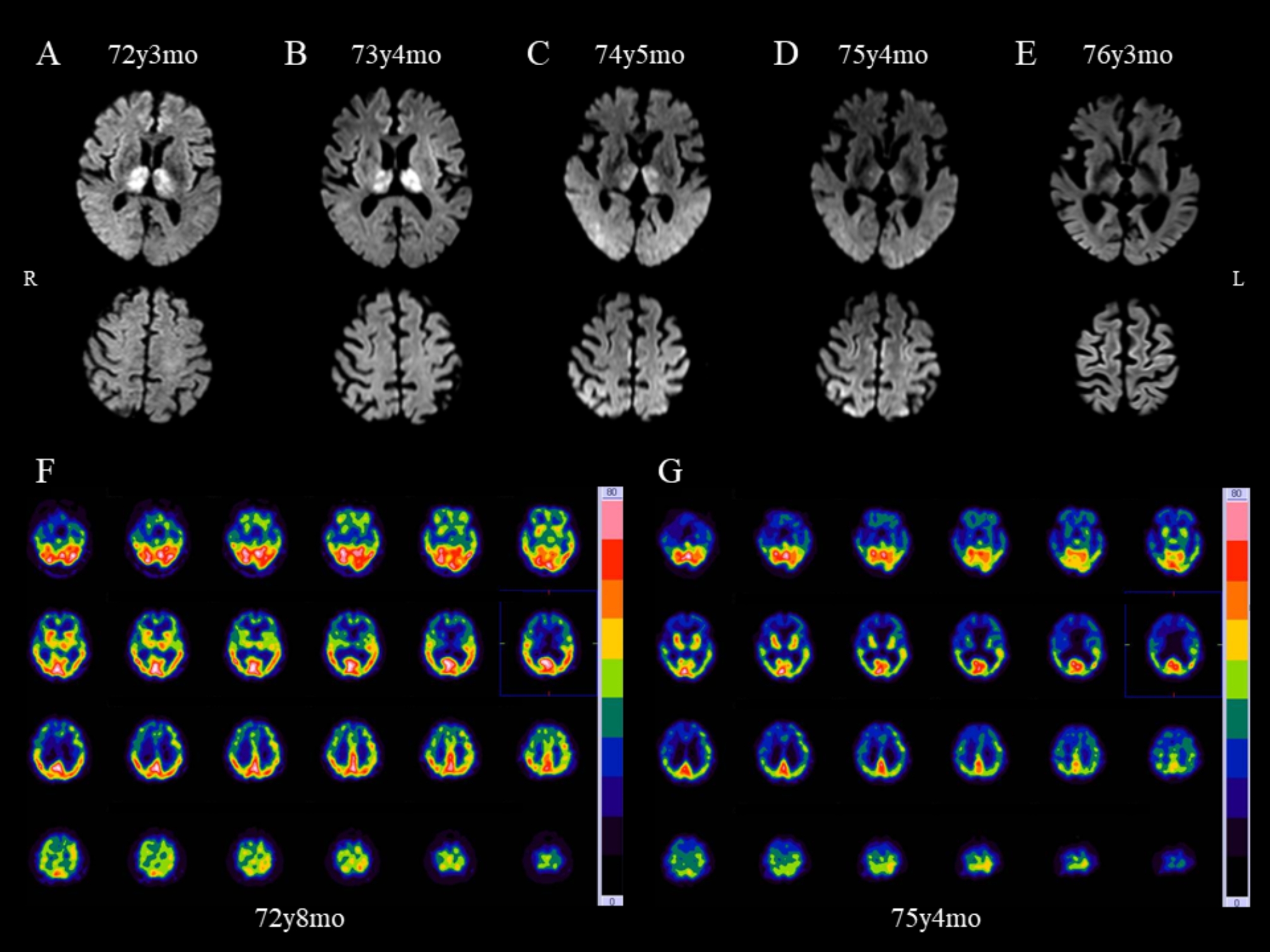
Fig. 1.

Brain imaging in an individual with Creutzfeldt–Jakob disease. Serial brain diffusion-weighted magnetic resonance imaging (DWI) scans (A–E) and technetium-99 m (99mTc)-ethylcysteinate dimer (ECD) single-photon emission computed tomography (SPECT) (F, G). Initial DWI performed 1 year after disease onset showed bilateral thalamic high signals with no cortical abnormalities (A). DWI conducted 2 years after disease onset exhibited findings similar to those in A (B). Three years after onset, DWI hyperintensities in the thalamus were less distinct and reduced in size (C upper); a few cortical lesions appeared (C lower). Four years (D) and 5 years (E) after onset, the thalamus showed atrophy and cortical hyperintensities became increasingly apparent over time. SPECT imaging in the early stage of the disease showed no thalamic hypoperfusion (F), whereas in the advanced stage, generalized hypoperfusion including in bilateral thalami was observed (G). The age of the individual is indicated on the images. y, years; mo, months
