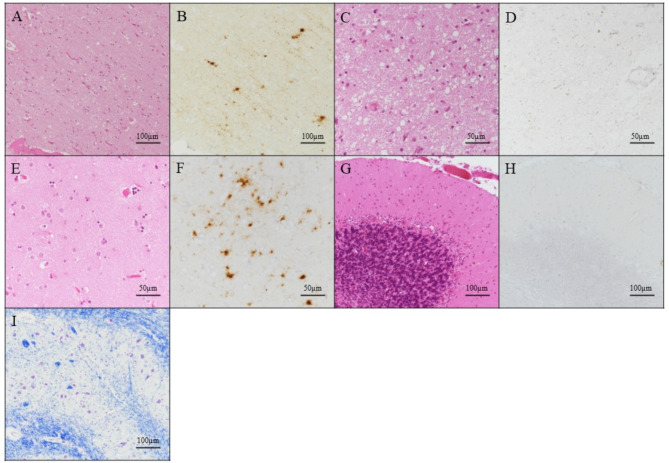
Fig. 3.
Postmortem brain pathology in an individual with Creutzfeldt–Jakob disease. Microscopic findings of the frontal lobe (A, B), anterior thalamic nucleus (C, D), caudate nucleus (E, F), cerebellum (G, H), and inferior olivary nucleus (I) with hematoxylin and eosin staining (A, C, E and G), anti-prion protein (PrP) immunostaining using 3F4 antibody (B, D, F and H), and Klüver–Barrera staining (I)

