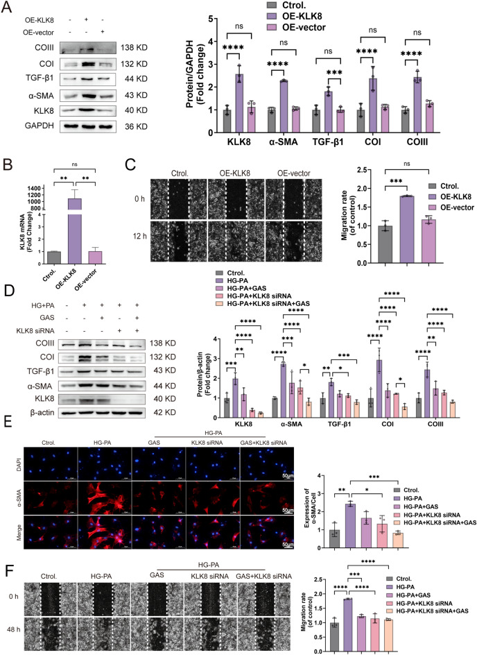
Fig. 5.
KLK8 mediates the differentiation, collagen synthesis, and migration of HG-PA-exposed CFs. A–C OE-KLK8-transfected CFs for 12 h. A OE-KLK8 increased the expression of the fibrosis-related proteins KLK8, α-SMA, TGFβ1, Collagen I, and Collagen III in CFs. B KLK8 mRNA was highly expressed in OE-KLK8-exposed CFs. C OE-KLK8-exposed CFs. The cell migration capacity was measured via a wound healing assay; scale bar = 50 μm. D–F CFs were transfected with KLK8 siRNA for 8 h, exposed to HG-PA, and then incubated for 40 h with or without GAS (5 µM). D Immunoblots showing the expression of the fibrosis-associated proteins KLK8, α-SMA, TGFβ1, Collagen I, and Collagen III. E Immunofluorescence staining of α-SMA is shown; α-SMA (green) and cell nuclei were restained with DAPI (blue); scale bar = 50 μm. F Wound healing assay; scale bar = 50 μm. The data are expressed as the mean ± SEM of three independent experiments; *p < 0.05, **p < 0.01, ***p < 0.001, and ****p < 0.0001.

