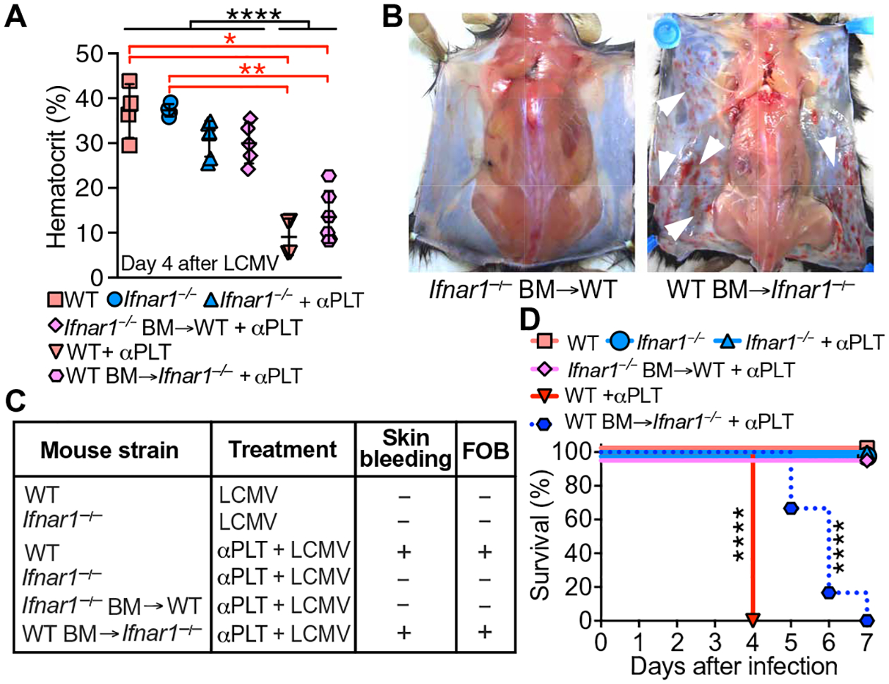
Fig. 2. Bleeding in LCMV-infected mice is mediated by IFNAR1 expressed on BM cells.

(A) WT mice (n = 4), Ifnar1−/− mice (n = 6), Ifnar1−/− mice with WT BM (n = 6), and WT mice with Ifnar1−/− BM (n = 5) were injected with αPLT followed by 5 × 10⁵–pfu LCMV. Controls (WT and Ifnar1−/−, n = 4 mice per group) received phosphate-buffered saline (PBS) instead of αPLT. Hematocrit in blood from the retro-orbital venous sinus was measured 4 days after infection. Data are shown as scatterplot with means ± SD. (B) Skin bleeding (white arrowheads) or lack thereof in chimeric mice. (C) Summary of the occurrence of skin bleeding and FOB. (D) Survival 7 days after infection without/with platelet depletion. N values for (C) and (D) are the same as in (A). Statistical analysis: (A) one-way analysis of variance (ANOVA) and Tukey’s posttest (black asterisks) or Kruskal-Wallis with Dunn’s posttest (red asterisks); (D) χ2 analysis with Mantel-Cox log-rank test. ****P < 0.0001; **P < 0.01; *P < 0.05.
