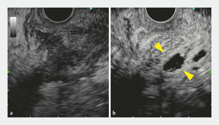
Fig. 2.
Endoscopic ultrasound images. a The initially targeted region. Despite the absence of an anechoic lesion, a mixed hypo- and hyperechoic area around the pancreas was observed under fundamental B-mode. b The initially targeted region was recognized as only minimal avascular areas (yellow arrowheads) on a contrast-enhanced harmonic image.

