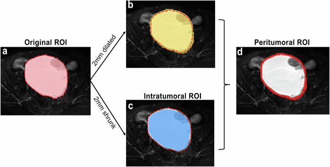
Fig. 3.
An example of the original, intratumoral, and peritumoral ROIs in a 64-year-old patient with left clear cell carcinoma. The “red dotted line” represents the tumor boundary. a The original ROI was the area within the tumor boundary; b 2 mm dilation of the tumor boundary; c the intratumoral ROI was derived from 2 mm shrinkage of the tumor boundary; d the peritumoral ROI was a 4 mm-thick ring, achieved by dilating the tumor boundary 2 mm outward and shrinking it 2 mm inward. ROI, region of interest

