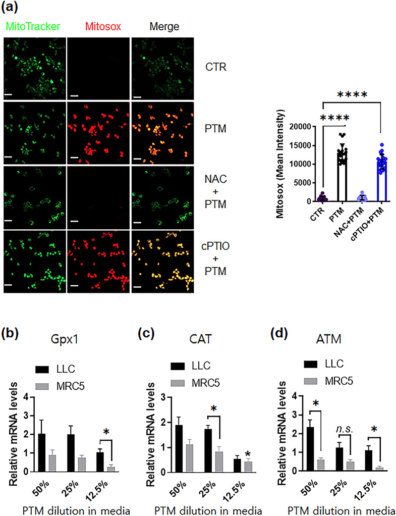
Fig. 3.
Plasma-treated medium detoxifies antioxidant enzymes in LLC or MRC5 cells. (a) (Left) Representative confocal microscopy images indicate MitoTracker (green) and Mitosox (red) staining of LLCs incubated in different conditions: the control, 25% PTM, and 25% PTM pre-incubated with 10 mM NAC or 0.1 mM cPTIO for 5 h; scale bar, 10 µM. (Right) The intensity of red fluorescence was quantified using Zen blue software; n = 20, 25, 19, and 27 single cells for control, PTM alone, and PTM incubation with pre-incubated NAC or cPTIO, respectively. The mean ± SED represents results from three independent experiments. Unpaired Student’s t-test was used to calculate statistical significance; ****p < 0.0001. (b-d) PTM stimulates anticancer signals in LLCs or normal lung MRC5 cells. The mRNA expression of Gpx1 (glutathione peroxidase), CAT (catalase), and ATM gene was quantified by qRT-PCR. Fold changes of the three target genes were normalized using the 18 S mRNA or GAPDH control. n = 3, mean ± sem. *p < 0.05, n.s., not significant

