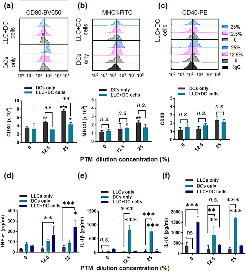
Fig. 6.
Plasma-treated medium (PTM) stimulates dendritic cells (DCs) alone and in a co-culture system. DCs were incubated alone (12.5 or 25% PTM) or co-cultured with Lewis lung cancer (LLC) cells in the presence of PTM. The DCs were cultured either alone or separated by a transwell system. After 24 h, surface marker expression was determined. (a-c). Representative histograms and bar plots demonstrate CD80-BV650, MHCII-FITC, and CD40-PE levels in the DCs only or in DCs with the co-culture, using flow cytometry, respectively. The data were analyzed and gated using FlowJo. The statistical significance of the difference between DC-alone and tumor cell co-culture groups was determined via a two-way ANOVA; *p < 0.05 and **p < 0.01; n.s., insignificant. Concurrently, data compared to the untreated cells using a two-tailed unpaired Student’s t-test are shown; *p < 0.05 and **p < 0.01, mean ± SEM; n = 4–5. (d-f) The TNF-α, IL-1β, and IL-10 levels in the supernatants from LLC cells, DCs cultured alone, DCs incubated with tumor cells, and both treated with 12.5% or 25% PTM, were measured. Comparison between LLCs, DCs, and LLC + DC cells using a two-way ANOVA; **p < 0.01, and ***p < 0.001. n.s., not significant. Statistical significance was determined using a two-tailed unpaired Student’s t-test; *p < 0.05, **p < 0.01, and ***p < 0.001; n = 4

