Correction to: Particle and Fibre Toxicology (2017) 14:13
10.1186/s12989-017-0193-5
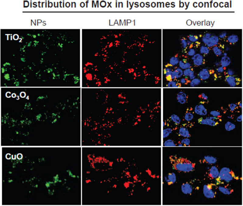
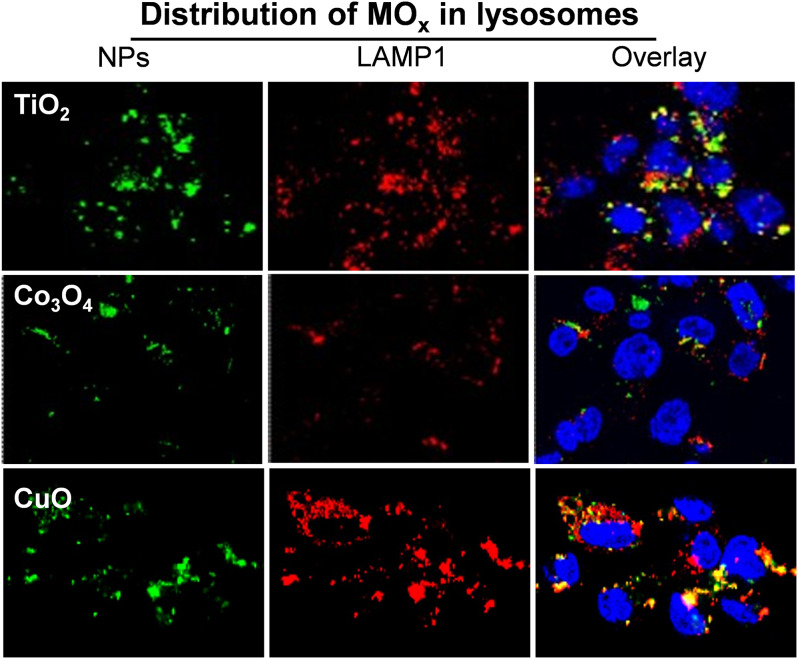
Following publication of the original article [1], the authors reported that the confocal images of TiO2 and Co3O4 in Figure S1 were erroneously uploaded. The corrected version is provided below. The error does not affect the conclusions of the study.
The incorrect version is:
Fig. S1.
Intracellular distribution of MOx by confocal imaging. THP-1 cells were exposed to 25 µg/mL TiO2, Co3O4 or CuO nanoparticle suspensions for 16 h. Then the cells were stained with Hoechst 33,342 and Alexa Fluor 594 labeled anti-LAMP1 to visualize the nuclei and lysosomes, respectively, by SP2 1P/FCS and Leica confocal SP2 MP-FLIM microscope
The correct version is:
Fig. S1.
Intracellular distribution of MOx by confocal imaging. THP-1 cells were exposed to 25 µg/mL TiO2, Co3O4 or CuO nanoparticle suspensions for 16 h. Then the cells were stained with Hoechst 33,342 and Alexa Fluor 594 labeled anti-LAMP1 to visualize the nuclei and lysosomes, respectively, by SP2 1P/FCS and Leica confocal SP2 MP-FLIM microscope
The original article [1] has been corrected.
Footnotes
The online version of the original article can be found at 10.1186/s12989-017-0193-5.
Publisher’s note
Springer Nature remains neutral with regard to jurisdictional claims in published maps and institutional affiliations.
Xiaoming Cai and Anson Lee contributed equally to this work.
Contributor Information
Tian Xia, Email: txia@ucla.edu.
Ruibin Li, Email: liruibin@suda.edu.cn.
Reference
- 1.Cai X, Lee A, Ji Z, et al. Reduction of pulmonary toxicity of metal oxide nanoparticles by phosphonate-based surface passivation. Part Fibre Toxicol. 2017;14:13. 10.1186/s12989-017-0193-5. [DOI] [PMC free article] [PubMed] [Google Scholar]


