Figure 2.
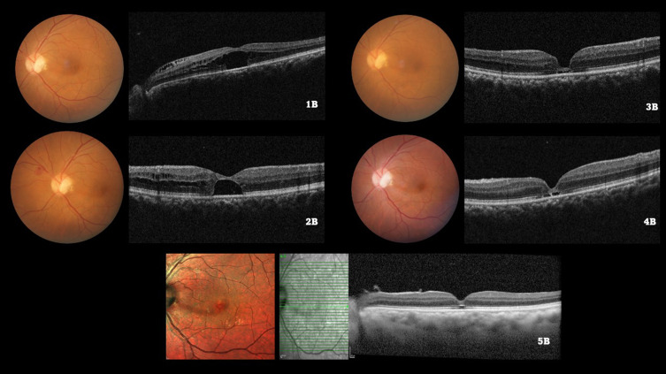
Color fundus photograph of the left eye of a 55/M illustrating the optic disc pit at baseline (1A) and subsequent visits (2A, 3A, 4A). The baseline spectral domain optical coherence tomography (1B) illustrates the serous macular detachment (SMD) along with the retinoschiatic lesions (RL). After undergoing a pars plana vitrectomy with internal limiting membrane (ILM) peeling and ILM plug and SF6 gas injection, the SMD and RL reduced by 3 months (2B). Subsequently, complete resolution of both the SMD and RL was observed by 6 months (3B), which was maintained upto months 12 (4B) and 24 (5B) respectively.

