
FIGURE 1.
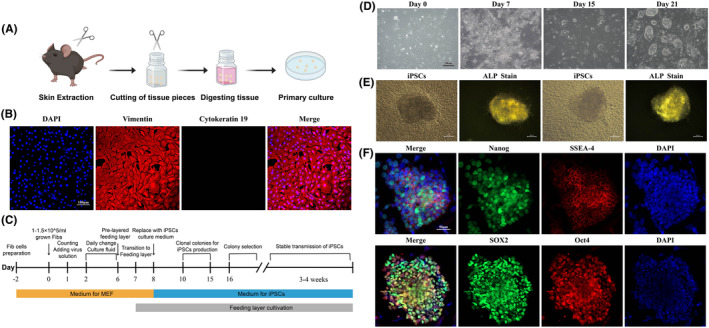
Acquisition of mouse Fibs and iPSCs construction. (A) Extraction and culture process of mouse Fibs; (B) Immunofluorescence of mouse Fibs showed high expression of vimentin and no expression of cytokeratin; (C) Flowchart of iPSCs construction; (D) Cell morphology at 0, 7, 15 and 21 d in iPSCs constructs; (E) ALP staining of iPSCs colonies showed high expression of ALP; (F) immunofluorescence of iPSCs.