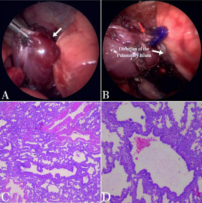
FIGURE 2.
(A) During the thoracoscopic examination, a solid mass approximately 30 mm × 25 mm × 10 mm in size, dark red in color, was found in the left posterior mediastinum. The mass was encapsulated by its own visceral pleura, had a smooth surface, was independent of the normal lung tissue, and was not connected to the normal bronchi (arrow). (B) A nourishing vessel about 3 mm in diameter was identified at the base of the lesion (red arrow), which was supplied by the pulmonary artery. (C,D) Histopathological examination (HE × 50 and HE × 100) showed bronchioles, alveoli, and alveolar structures within the tissue. Bronchiectasis was covered with pseudostratified fibrous columnar epithelium, surrounded by fibromuscular tube walls. Cartilage plates were observed in some bronchial walls, consistent with extralobar pulmonary sequestration.

